

 paper
paperInformation
- Publication Type: Master Thesis
- Workgroup(s)/Project(s):
- Date: July 2010
- TU Wien Library:
- Diploma Examination: 25. July 2010
- First Supervisor:
- Milos Sramek
- Andrej Varchola
Abstract
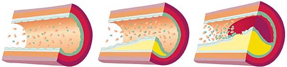
Automated processing and visualization of vascular structures is a common task in medical imaging. Maximum Intensity Projection (MIP) and Curved Planar Reformation (CPR) are well established and robust methods for clinical use. In case of calcified vessel walls, occlusion prevents exploring the inside of the vessels when using MIP. CPR allows to cut a single vessel along its centerline and to visualize the lumen. Extending the idea of CPR, a novel automatic method for vessel visualization is proposed. It works with multiple vessel centerlines that do not necessarily need to be connected into a tree structure. Arbitrarily complex vascular structures are rendered in the volume as point sets and optionally, occlusion halos are created around them to enhance depth perception. Vessel centerlines are automatically extracted from a volumetric data-set after performing feature extraction in a scale-space. The user is provided with the ability to control the final image and he or she can visually select the desired centerlines with visual queries by stroking with the mouse. Furthermore, a combination with the recent Maximum Intensity Difference Accumulation (MIDA) visualization technique is presented, which has the advantages of Direct Volume Rendering (DVR) such as occlusion and depth cues, but does not require an explicit transfer function specification. It is demonstrated how the proposed technique can be applied to large data-sets, particularly to data featuring peripheral arterial occlusive diseases or in order to detect possible embolisms as presented on a pulmonary data-set.Additional Files and Images

Weblinks
No further information available.BibTeX
@mastersthesis{mistelbauer-2010-pvv,
  title =      "Automated Processing and Visualization of Vessel Trees",
  author =     "Gabriel Mistelbauer",
  year =       "2010",
  abstract =   "Automated processing and visualization of vascular
               structures is a common task in medical imaging. Maximum
               Intensity Projection (MIP) and Curved Planar Reformation
               (CPR) are well established and robust methods for clinical
               use. In case of calcified vessel walls, occlusion prevents
               exploring the inside of the vessels when using MIP. CPR
               allows to cut a single vessel along its centerline and to
               visualize the lumen. Extending the idea of CPR, a novel
               automatic method for vessel visualization is proposed. It
               works with multiple vessel centerlines that do not
               necessarily need to be connected into a tree structure.
               Arbitrarily complex vascular structures are rendered in the
               volume as point sets and optionally, occlusion halos are
               created around them to enhance depth perception. Vessel
               centerlines are automatically extracted from a volumetric
               data-set after performing feature extraction in a
               scale-space. The user is provided with the ability to
               control the final image and he or she can visually select
               the desired centerlines with visual queries by stroking with
               the mouse. Furthermore, a combination with the recent
               Maximum Intensity Difference Accumulation (MIDA)
               visualization technique is presented, which has the
               advantages of Direct Volume Rendering (DVR) such as
               occlusion and depth cues, but does not require an explicit
               transfer function specification. It is demonstrated how the
               proposed technique can be applied to large data-sets,
               particularly to data featuring peripheral arterial occlusive
               diseases or in order to detect possible embolisms as
               presented on a pulmonary data-set.",
  month =      jul,
  address =    "Favoritenstrasse 9-11/E193-02, A-1040 Vienna, Austria",
  school =     "Institute of Computer Graphics and Algorithms, Vienna
               University of Technology ",
  URL =        "https://www.cg.tuwien.ac.at/research/publications/2010/mistelbauer-2010-pvv/",
}