

Information
- Publication Type: Bachelor Thesis
- Workgroup(s)/Project(s):
- Date: November 2017
- Date (Start): 10. June 2017
- Date (End): 28. November 2017
- Matrikelnummer: 01325652
- First Supervisor: Eduard Gröller

Abstract
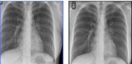
In this thesis different state-of-the-art machine learning frameworks were implemented and evaluated on chest radiographs to classify them into tuberculotic or healthy radiographs. Traditional explicit feature engineering was performed, as well as different deep learning approaches were applied. For the deep learning experiments different publicly available architectures were compared in two different tasks. The first task with deep learning was to use a Convolutional Neural Network, already trained on a different task, to extract features of the chest radiographs. These features were then classified separately. The second experiment was to use a Convolutional Neural Network, again pretrained on a different task, and train this network carefully again on the chest radiographs. The results of the different frameworks were summarized, evaluated and presented in tables.Additional Files and Images

Weblinks
No further information available.BibTeX
@bachelorsthesis{unger_2017,
  title =      "Evaluation of Machine Learning Frameworks on Tuberculosis
               Classification of Chest Radiographs",
  author =     "Katharina Unger",
  year =       "2017",
  abstract =   "In this thesis different state-of-the-art machine learning
               frameworks were implemented and evaluated on chest
               radiographs to classify them into tuberculotic or healthy
               radiographs. Traditional explicit feature engineering was
               performed, as well as different deep learning approaches
               were applied. For the deep learning experiments different
               publicly available architectures were compared in two
               different tasks. The first task with deep learning was to
               use a Convolutional Neural Network, already trained on a
               different task, to extract features of the chest
               radiographs. These features were then classified separately.
               The second experiment was to use a Convolutional Neural
               Network, again pretrained on a different task, and train
               this network carefully again on the chest radiographs. The
               results of the different frameworks were summarized,
               evaluated and presented in tables.",
  month =      nov,
  address =    "Favoritenstrasse 9-11/E193-02, A-1040 Vienna, Austria",
  school =     "Institute of Computer Graphics and Algorithms, Vienna
               University of Technology ",
  URL =        "https://www.cg.tuwien.ac.at/research/publications/2017/unger_2017/",
}