

 Master thesis
 Poster
Information
- Publication Type: Master Thesis
- Workgroup(s)/Project(s):
- Date: August 2019
- Date (Start): 5. May 2018
- Date (End): 5. August 2019
- TU Wien Library:
- Open Access: yes
- First Supervisor: Eduard Gröller

Abstract
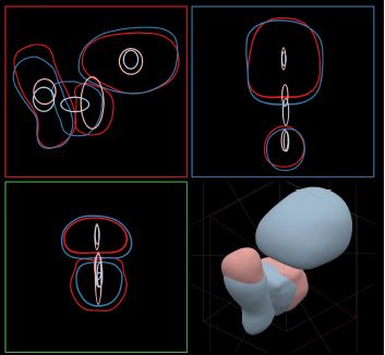
Pelvic organs such as the bladder, rectum or prostate have highly variable shapes that change over time, due to their soft and flexible tissue and varying filling. Recent clinical work suggests that these variations might affect the effectiveness of radiation therapy treatment in patients with prostate cancer. Although in clinical practice small correction steps are performed to re-align the treated region if the organs are shifted, a more in-depth understanding and modeling might prove beneficial for the adaptation of the employed treatment planning strategy. To evaluate the viability and to account for the variability in the population of certain treatment strategies, cohort studies are performed analyzing the shape and position variability of pelvic organs. In this thesis, we propose a web-based tool that is able to analyze a cohort of pelvic organs from 24 patients across 13 treatment instances. Hereby we have two goals: On the one hand, we want to support medical researchers analyzing large groups of patients for their shape variability and the possible correlations to side effects. On the other hand, we want to provide support for medical experts performing individual patient treatment planning. Our tool offers both the option to analyze a large cohort of different organ shapes, by first modeling them in a shape space and then analyzing the shape variations on a per-patient basis. While this first part aims at providing users with an overview of the data, we also give them the option to perform a detailed shape analysis, where we highlight the statistically aggregated shape of a patient or a specified group using a contour variability plot. Finally, we demonstrate several possible usage scenarios for our tool and perform an informal evaluation with two medical experts. Our tool is the first significant step in supporting medical experts in demonstrating the need for adaptation in radiation therapy treatments to account for shape variability.Additional Files and Images


Weblinks
BibTeX
@mastersthesis{Grossmann_MA,
  title =      "Pelvis Runner - Comparative Visualization of Anatomical
               Changes",
  author =     "Nicolas Grossmann",
  year =       "2019",
  abstract =   "Pelvic organs such as the bladder, rectum or prostate have
               highly variable shapes that change over time, due to their
               soft and flexible tissue and varying filling. Recent
               clinical work suggests that these variations might affect
               the effectiveness of radiation therapy treatment in patients
               with prostate cancer. Although in clinical practice small
               correction steps are performed to re-align the treated
               region if the organs are shifted, a more in-depth
               understanding and modeling might prove beneficial for the
               adaptation of the employed treatment planning strategy. To
               evaluate the viability and to account for the variability in
               the population of certain treatment strategies, cohort
               studies are performed analyzing the shape and position
               variability of pelvic organs. In this thesis, we propose a
               web-based tool that is able to analyze a cohort of pelvic
               organs from 24 patients across 13 treatment instances.
               Hereby we have two goals: On the one hand, we want to
               support medical researchers analyzing large groups of
               patients for their shape variability and the possible
               correlations to side effects. On the other hand, we want to
               provide support for medical experts performing individual
               patient treatment planning. Our tool offers both the option
               to analyze a large cohort of different organ shapes, by
               first modeling them in a shape space and then analyzing the
               shape variations on a per-patient basis. While this first
               part aims at providing users with an overview of the data,
               we also give them the option to perform a detailed shape
               analysis, where we highlight the statistically aggregated
               shape of a patient or a specified group using a contour
               variability plot. Finally, we demonstrate several possible
               usage scenarios for our tool and perform an informal
               evaluation with two medical experts. Our tool is the first
               significant step in supporting medical experts in
               demonstrating the need for adaptation in radiation therapy
               treatments to account for shape variability.",
  month =      aug,
  address =    "Favoritenstrasse 9-11/E193-02, A-1040 Vienna, Austria",
  school =     "Research Unit of Computer Graphics, Institute of Visual
               Computing and Human-Centered Technology, Faculty of
               Informatics, TU Wien",
  URL =        "https://www.cg.tuwien.ac.at/research/publications/2019/Grossmann_MA/",
}